


NEXTSCREEN
is a joint Doctoral Network supported by the Marie Skłodowska-Curie Actions (MSCA) of Horizon Europe, that will recruit and train 10 PhD students in the fields of biophotonics, micromanipulation, machine learning and in vitro diagnostics.
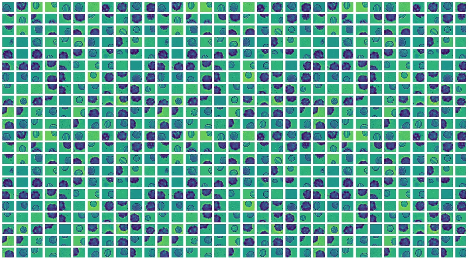
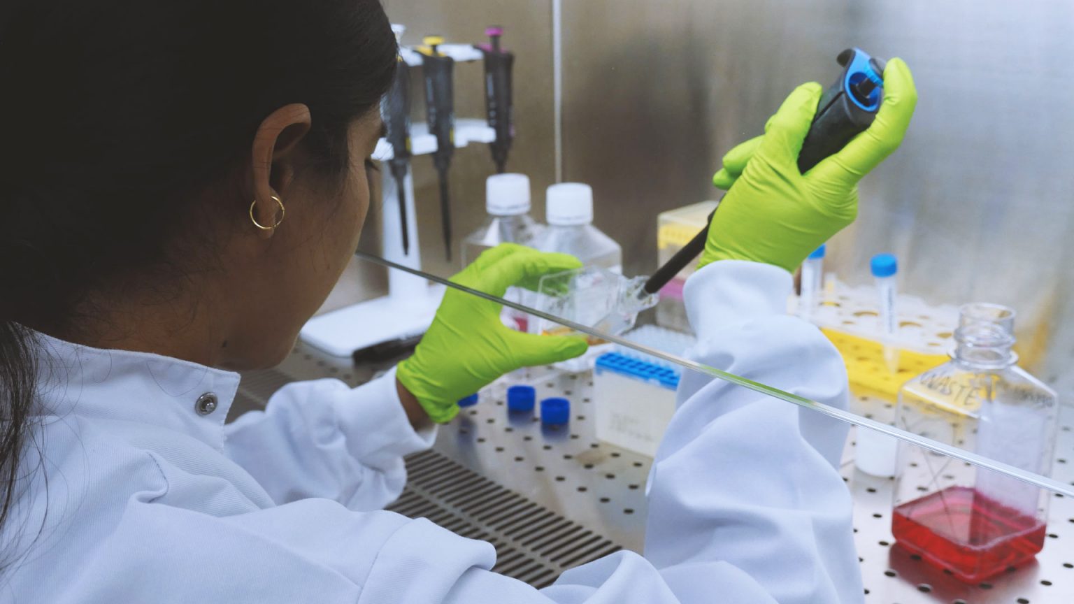
Using these technologies, they will characterize cells and bioparticles, screening large cellular populations. The ultimate goal is to initiate the development of diagnostics tools, that could be adopted in clinical settings on a large scale.
The project brings together research groups, SMEs and large companies in Italy, France, Spain, Germany, Greece, Switzerland, with complementary know-how in high-resolution microscopy, high-precision microfluidics, biotechnologies, and deep-learning, that will facilitate the development of data-driven cells and bioparticle screening.
We are looking for 10 candidates with background (Master level) in one of the following fields: physics, engineering, computer science, biotechnology, biomedicine. The consortium includes academic and industrial partners and dedicated training tracks will be identified for each of the 10 candidates, in order to develop their cross disciplinary skills and international networking.
The participants will take part of an international and transdisciplinary network, in top research centers and industries in Europe, equipped with state-of-the-art technologies, and will benefit of highly competitive salaries.
The goal of the project is to boost their career in optical engineering, material sciences and micromanufacturing, computer sciences and biotechnologies.
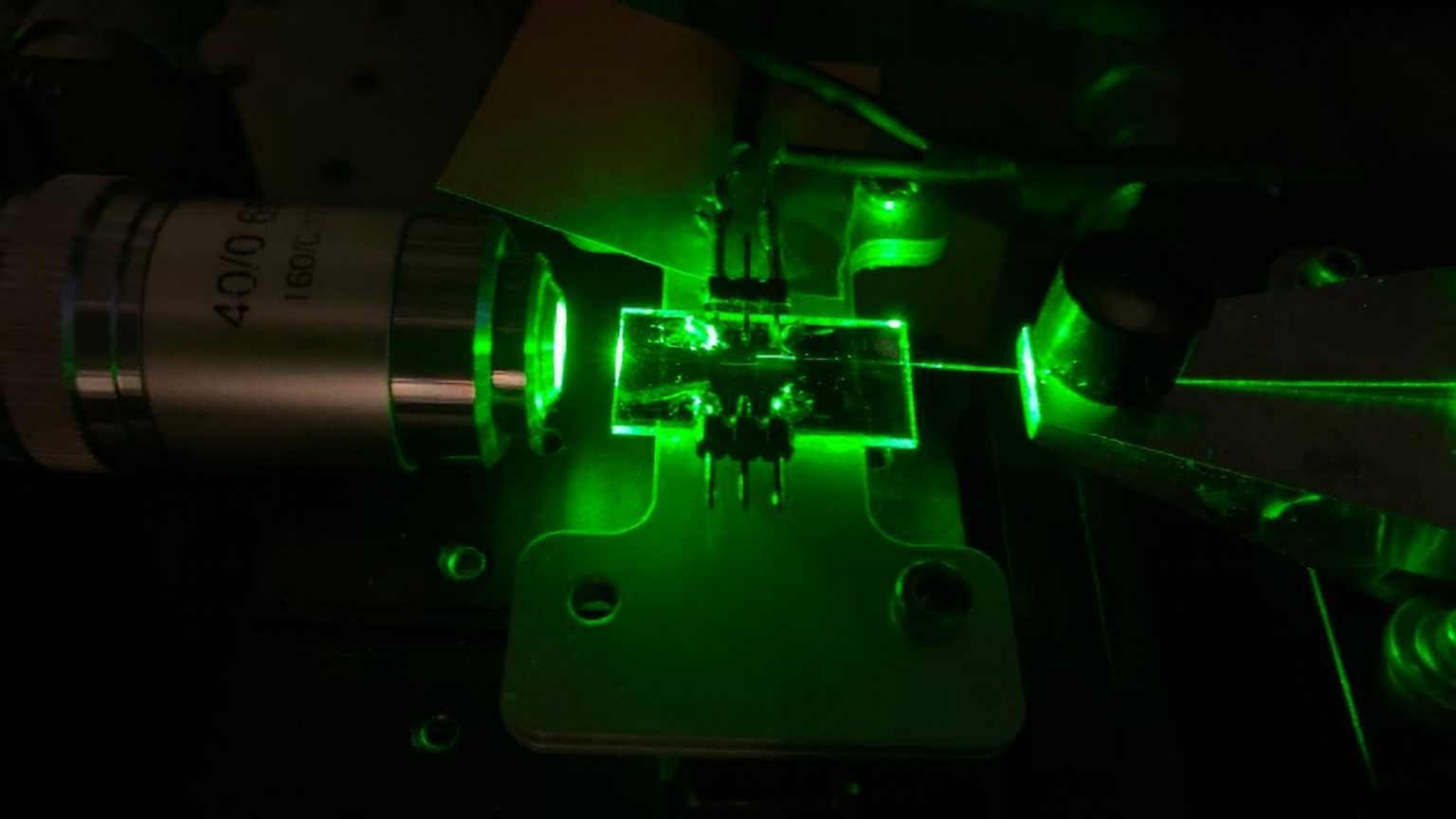
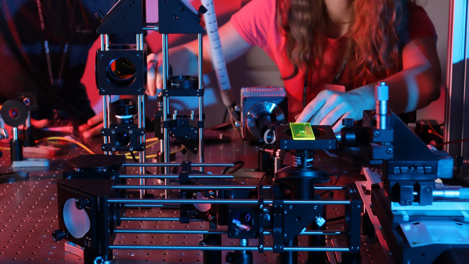
The PhD students will work together on imaging flow cytometry, an imaging technology that combines the subcellular resolution of optical microscopy with the high-content-screening capability of flow cytometry.
Several projects will be carried out by the PhD students with the goal to reduce the cost and complexity of imaging flow cytometry, empower it with novel contrast mechanisms, build high-resolution automatic microscopes at the diffraction limit and beyond; develop real-time data processing tools able to detect and recognize the samples, circumventing the need for manual annotation.
Existing technology
Flow cytometry is an established technology for detection, characterization, and sorting of populations of cells and bioparticles, widely adopted in cellular biology and biotechnology. The samples (derived from cell cultures, liquid biopsies, or disaggregated tissues) move along a liquid stream and are automatically analysed while passing through a laser beam. Light scattering, absorption and fluorescence at multiple wavelengths, are some of the acquired parameters. Imaging Flow Cytometry (IFC) adds spatial resolution as an additional measurement parameter, capturing microscopy images of the samples that move along the stream.
Imaging is a key parameter in studying cell morphology, cytotoxicity, drugs uptake, protein–protein interactions, co-localization, which have been widely used in biological and pharmaceutical studies.
The consortium includes academic and industrial partners that are leading the field of imaging flow cytometry. State of the art technologies have been recently developed by the partners (see the figures for some examples) opening the way to three dimensional and high-throughput imaging. The goal is to further enhance these method and open new applications in automatic biomedical screening.
Objectives
- To lower the cost and complexity of automatic cellular and particle screening, maintaining high resolution and throughput. The specific objectives are to: (i) develop an optofluidic lab-on-chip, that integrates the illumination, the sample scanning and the detection components of IFC in a single mm-scaled device, removing the need for periodic optical alignment and refocusing; (ii) identify low-cost materials for the fabrication of compact and scalable imaging/fluidics probes, suitable for high-resolution biomedical screening.
- To extend the capabilities of imaging flow cytometry with novel contrast mechanisms and higher resolution. The specific objectives are to develop new image flow cytometry modalities for blood derived samples including: (i) three-dimensional label-free acquisition at high rate; (ii) deformability acquisition in high throughput; (iii) high resolution imaging at, and beyond, the diffraction limit.
- To demonstrate the blood-cell classification capability of deep learning, avoiding manual annotation in imaging flow cytometry. The specific objectives are to: (i) create an open-source platform for objects detection, segmentation and labelling for high-throughput IFC; (ii) develop weakly supervised deep learning methods for the classification of blood derived samples, that circumvent the need for manual labelling.
- To introduce a protocol for quality control, reporting, and reproducibility in imaging flow cytometry. The specific objectives are to: (i) conceive and prepare a standard test sample for IFC; (ii) identify a list of guidelines for open data acquisition, processing and reporting.
To adopt imaging flow cytometry for identifying and characterizing tumour biomarkers in blood derived samples. The specific objectives are to: (i) demonstrate the detectability of known and new tumour biomarkers, screening biological nanoparticles and cells derived from blood in high throughput. (ii) investigate the abundance and (co)localization of antigens, along with the blood-cells morphology and deformability, as markers of disease. The long-term goal is to devise in-vitro diagnostics devices for liquid biopsies that can be used by non-technical users in clinical settings.
News
Applications are open for the International School on Shaping Light in Space & Time for Biomedical Imaging. Join us in stunning Erice, Sicily, May 14–20, 2025. Please share with students & young researchers!
More info: https://www.in2sight.eu/?page_id=1566 #BiomedicalImaging #Microscopy
Great first workshop with the NEXTSCREEN consortium! Thanks to @ICFOnians and @SLN_ICFOfor organizing the hands-on microscopy lab training for our PhD students. Best of luck to all the students as they kick off their research!
4Contact us
Project coordinator: Andrea Bassi
Project manager: Giorgia Lancerotti
Webmaster: Maurizio Contran
